
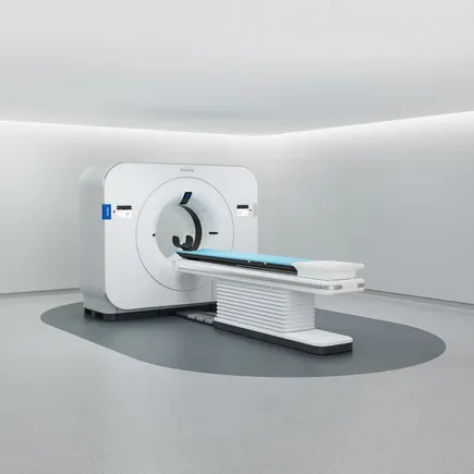
The medical technology landscape has reached a significant milestone with the United States Food and Drug Administration granting 510(k) clearance to Philips’ Verida, a revolutionary detector-based spectral Computed Tomography (CT) system integrated with advanced artificial intelligence. This regulatory approval marks a pivotal moment for Royal Philips as it seeks to aggressively expand its presence in the premium diagnostic imaging sector, setting a strategic target to capture a larger portion of the global market through 2028. By combining the physical advantages of spectral energy detection with the computational power of deep learning AI, the Verida system represents a shift in how clinicians approach complex diagnostic challenges, prioritizing both high-speed throughput and precision medicine.
The Evolution of Spectral CT and the Verida Innovation
To understand the significance of the Verida system, one must first examine the fundamental differences between conventional CT imaging and spectral CT. Traditional CT scanners utilize a single energy X-ray beam to create cross-sectional images of the body. While effective, these systems often struggle to differentiate between various materials that have similar densities, such as certain types of soft tissue, calcifications, and contrast agents.
Spectral CT, also known as dual-energy CT, solves this by measuring how tissues absorb different levels of X-ray energy. This allows the system to characterize the chemical composition of tissues and fluids, effectively "unmasking" details that would otherwise remain hidden. Philips’ Verida system takes this a step further by utilizing a specialized detector-based approach. Unlike older methods that required scanning a patient twice or switching energy levels rapidly—which could lead to motion blur or increased radiation—the Verida’s detector captures spectral data simultaneously during every scan. This ensures that spectral information is available for every patient, every time, without the need for special protocols.
The integration of artificial intelligence is the second pillar of the Verida’s design. AI algorithms are utilized throughout the imaging chain, from initial scan planning to the reconstruction of the final images. This synergy between hardware and software allows the system to achieve levels of clarity and efficiency that were previously considered unattainable in high-volume clinical settings.
Enhancing Clinical Throughput and Operational Efficiency
One of the most pressing challenges facing modern radiology departments is the sheer volume of patients. With an aging global population and an increasing reliance on diagnostic imaging for chronic disease management, hospitals are under immense pressure to increase "throughput"—the number of patients scanned per day—without sacrificing diagnostic quality.
Philips has engineered the Verida system specifically to address these operational bottlenecks. The system is capable of reconstructing 145 images per second, a feat that allows healthcare professionals to view completed exams in as little as 30 seconds. According to data released by Philips, the Verida is twice as fast as its predecessor, the IQon Spectral CT. For a high-capacity medical facility operating on a 16-hour workday, this speed translates into the ability to perform up to 270 exams per day.
This efficiency does not merely benefit the hospital’s bottom line; it has a direct impact on patient care. Faster reconstruction times mean that radiologists can provide answers to emergency department physicians more quickly, potentially saving lives in "stroke code" or trauma situations where every second counts. Furthermore, the high-speed workflow reduces the time patients spend on the scanning table, improving the overall patient experience and reducing the likelihood of movement-related image artifacts.
The Role of AI in Radiation Safety and Diagnostic Precision
Radiation safety remains a primary concern in the field of medical imaging. The Verida system addresses this through its AI-driven reconstruction engines. At a recent investor event, Philips executive Frans van Houten and Chief Business Leader for Precision Diagnosis, Bert van Meurs, along with other key leaders like Xue, highlighted that the system’s AI enables an 80% reduction in radiation dose and image noise.
This reduction is achieved through sophisticated noise-cancellation algorithms that can distinguish between "true" anatomical data and electronic "noise" inherent in high-speed scanning. By cleaning up the image, the AI allows the system to use a lower dose of X-rays while still producing a diagnostic-quality image. This is particularly beneficial for pediatric patients and those requiring frequent follow-up scans, such as oncology patients.
Beyond safety, the AI enhances the system’s ability to detect subtle differences in tissue. For instance, in oncology, the ability to accurately quantify iodine uptake in a tumor can help clinicians determine whether a treatment is working long before the tumor actually changes in size. In cardiology, the spectral data can help differentiate between "soft" vulnerable plaques and stable calcified plaques in the coronary arteries, providing a much more nuanced view of a patient’s cardiovascular risk.
Strategic Market Positioning and the 2028 Roadmap
The FDA clearance of Verida is a cornerstone of Philips’ broader business strategy. The company has explicitly stated its goal to increase its share of the premium CT market over the next four years. This sector of the market is characterized by high-margin, high-performance systems purchased by major academic medical centers and large hospital networks.
The launch follows a successful rollout in Europe, where the Verida system received its CE Mark last year. The first global installation was initiated at a prominent clinical site in Madrid, Spain, in early 2024. With the U.S. market now open, Philips is positioned to compete for the large-scale replacement cycles currently occurring in American hospitals.
The financial implications are significant. The Verida system is priced between 1 million and 2 million euros (approximately $1.1 million to $2.2 million USD), depending on the specific configuration and regional market factors. This pricing strategy places the Verida firmly in the premium category, yet Philips argues that it offers a superior return on investment compared to emerging experimental technologies.
The Competitive Landscape: Spectral vs. Photon Counting
The diagnostic imaging market is currently witnessing a technological arms race. While Philips is doubling down on detector-based spectral CT, its primary competitors, GE HealthCare and Siemens Healthineers, are heavily invested in Photon-Counting CT (PCCT).
Siemens Healthineers was the first to market with a PCCT device, receiving FDA clearance for its Naeotom Alpha in 2021. GE HealthCare followed suit, securing clearance for its own photon-counting technology in March 2024. Photon-counting is widely regarded as the "holy grail" of CT imaging because it counts individual X-ray photons, providing even higher spatial resolution and better contrast-to-noise ratios than traditional spectral CT.
However, Philips has taken a pragmatic stance on this competition. During a February investor briefing, Philips executive Xue acknowledged the power of photon counting but argued that the technology is not yet optimized for high-throughput clinical environments. The primary barriers cited were cost and complexity.
According to Philips’ analysis, a photon-counting system can cost upwards of 3 million euros—roughly double the price of a Verida system. Furthermore, the maintenance and operational requirements for PCCT are currently higher, which may limit its utility in busy community hospitals. Philips’ strategy is to provide a "production-ready" spectral solution that offers most of the benefits of advanced imaging at a price point and speed that makes sense for the majority of healthcare providers today. The company has indicated it will enter the photon-counting market only when the technology has matured sufficiently to meet these high-volume clinical demands.
Implications for the Future of Radiology
The introduction of the Verida system reflects a broader trend in healthcare toward the "Quadruple Aim": improving the patient experience, improving the health of populations, reducing the per capita cost of healthcare, and improving the work life of healthcare providers.
For radiologists, the AI-powered spectral workflow reduces the cognitive load required to interpret complex cases. By providing automated spectral maps and "virtual non-contrast" images, the system simplifies the diagnostic process. For hospital administrators, the increased throughput and lower dose profile make the Verida an attractive asset for meeting regulatory requirements and managing rising patient volumes.
As Philips moves toward its 2028 targets, the success of the Verida will likely depend on how well it can bridge the gap between "standard" CT and the ultra-premium photon-counting systems. By positioning Verida as the most efficient and cost-effective path to advanced spectral diagnostics, Philips is betting that the market will prioritize clinical productivity and proven AI integration over the experimental potential of more expensive alternatives.
The FDA clearance of Verida serves as a clear signal that Philips remains a formidable player in the medical imaging space. With the global CT market projected to continue its steady growth, the integration of AI and spectral detection is no longer just a luxury for research institutions; it is becoming the new standard for diagnostic excellence in mainstream medicine. The coming years will reveal whether this "efficiency-first" approach will allow Philips to reclaim its dominance in the highly competitive premium imaging landscape.
